
“ FIVE -POINT MANAGEMENT ALGORITHM ”
The cornea is the clear, dome-shaped window at the front of your eye. It focuses light into your eye. Keratoconus is when the cornea thins out and bulges outward like a cone. Changing the shape of the cornea brings light rays out of focus. Its cause is unknown.Symptoms first appear during puberty or the late teens and include blurred vision and sensitivity to light and glare.Vision can be corrected with glasses or contact lenses early on. As condition progress, vision gets blurry and distorted, making daily tasks like reading, driving difficult.Advanced cases may require a cornea transplant.
By definition, Keratoconus is a slowly progressive, noninflammatory ectatic corneal disease characterized by changes in corneal collagen structure and organization. There is thinning of the corneal stroma that may or may not lead to irregular astigmatism with an associated decrease in visual acuity.
1. DEMOGRAPHICPARAMETERS :
It affects the rate of progression of the disease.
2. CONTACT LENS/SPECTACLES :
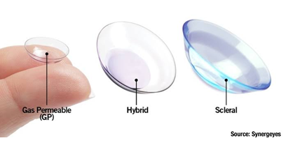
Common methods of vision correction includes spectacles, rigid gas permeable contact lenses specialized lenses - Piggy bank, Rose-K or Boston scleral lenses.
3. CORNEAL COLLAGEN CROSS-LINKING :
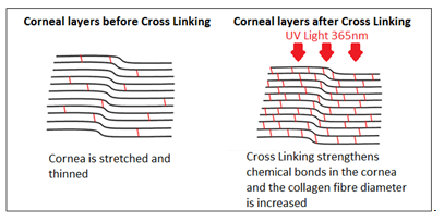
It is effective in stabilizing the progression of the disease. Intra corneal ring segments can improve vision by flattening the cornea in patients with mild to moderate keratoconus.
4. TOPOGRAPHY GUIDED-PHOTOREFRACTIVE KERATECTOMY/INTACTS :
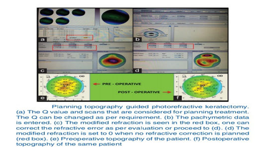
Topography‑guided custom ablation treatment betters the quality of vision by correcting the refractive error and improving the contact lens fit.
5. SURGICAL MANAGEMENT :
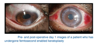
In advanced keratoconus with corneal scarring, lamellar or full thickness penetrating keratoplasty will be the treatment of choice.
By Dr Sukriti Gupta
JR Ophthalmology Department
Santosh Medical College & University